

Evaluation of Osteoblast and Fibroblast Viability on Bovine Amniotic Membrane Tropical Journal of Natural Product Research
Article Sidebar
Main Article Content
Abstract
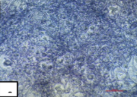
Guided tissue regeneration (GTR), as one of the periodontal treatments, is a reconstructive periodontal surgical technique where scaffold is an important element. Bovine amniotic membrane (BAM) has anti-inflammatory and antibacterial properties, also multiple growth factors which are useful for periodontal healing and regeneration processes. As a biomaterial with GTR potential, BAM must meet the criteria for membrane barriers, including biocompatibility. This study aimed to determine the viability of osteoblast and fibroblast cells following the application of bovine amnion membranes. The study used a design featuring a laboratory experiment with a post-test-only control group. The cells were divided into five groups, each with seven replicates. Osteoblast and fibroblast cells were treated with BAM for 24 hours, after which the viability of the osteoblast and fibroblast cells was determined using the microculture tetrazolium assay. Consequently, after the intervention, osteoblast and fibroblast cells vitality was 91.73% and 98.4%, respectively. The statistical analysis indicated no significant difference between the cell control and treatment groups. This finding demonstrates that post-administration of BAM, osteoblast and fibroblast cells displayed elevated cell viability, with a live cell percentage above 50%. This indicated that BAM is safe and non-toxic to osteoblast and fibroblast cells.
Downloads
Article Details

This work is licensed under a Creative Commons Attribution-NonCommercial-NoDerivatives 4.0 International License.
How to Cite
References
Kiany F and Moloudi F. Amnion membrane as a novel barrier in the treatment of intrabony defects: a controlled clinical trial. Int J Oral Maxillofac Implants. 2015; 30(3):639-647. Doi: 10.11607/jomi.3590
Min S, Yoon JY, Park SY, Kwon HH, Suh DH. Clinical effect of bovine amniotic membrane and hydrocolloid on wound by laser treatment: Prospective comparative randomized clinical trial. Wound Repair Regen. 2014; 22(2):212–219. Doi: 10.1111/wrr.12145
Caballe-Serrano J, Munar-Frau A, Ortiz-Puigpelat O, Soto-Penaloza D, Penarrocha M, Hernandez-Alfaro F. On the search of the ideal barrier membrane for guided bone regeneration. J Clin Exp Dent. 2018; 8(2):477-483. Doi: 10.4317/jced.54767
Faadhila TI, Valentina MN, Munadziroh E, Nirwana I, Soekartono H, Surboyo MDC. Bovine sponge amnion stimulates socket healing: A histological analysis. J Adv Pharm Technol Res. 2021; 12(1):99–103. Doi: 10.4103/japtr.JAPTR_128_20
Elkhenany H, El-Derby A, Elkodous MA, Salah RA, Lotfy A, El-Badri N. Applications of the amniotic membrane in tissue engineering and regeneration: the hundred-year challenge. Stem Cell Res Ther. 2022; 13(8):1-19. Doi: 10.1186/s13287-021-02684-0
Octarina, Munadziroh E, Razak FA. Physical Modification of Bovine Amniotic Membrane for Dental Application. J Int Dent Med Res. 2022; 14 (4):1425–1428.
Octarina, and Elly M. Characterization of Fresh Bovine Amnion Membrane Combined with Hydroxyapatite as Candidate Scaffold for Alveolar Bone Tissue Engineering. J Dent Indones. 2024; 31(3):208-214. Doi: 10.14693/jdi.v31i3.1722
Majzoub J, Barootchi S, Tavelli L, Wang CW, Chan HL, Wang HL. Guided tissue regeneration combined with bone allograft in infrabony defects: Clinical outcomes and assessment of prognostic factors. J Periodontol. 2020; 91(6):746–755. Doi: 10.1002/JPER.19-0336
Fillingham Y and Jacobs J. Bone grafts and their substitutes. The Bone Joint J. 2016; 98-B(1 Suppl A):6–9. Doi: 10.1302/0301-620X.98B.36350
Smith PC, Martínez C, Martínez J, McCulloch CA. Role of Fibroblast Populations in Periodontal Wound Healing and Tissue Remodeling. Front Physiol. 2019; 10(270):1-11. Doi: 10.3389/fphys.2019.00270
Ariestania V, Hendrijantini N, Prahasanti C, Prasetyo E, Kuntjoro M, Sari RP, Maharani AD. Cytotoxicity of HA-TCP Scaffold on Human Umbilical Cord Mesenchymal Stem Cells using MTT Assay. Int J Integr Engr. 2022; 14(2):1-6. Doi: 10.30880/ijie.2022.14.02.012
Apriasari ML, Nadia H, Hanifa N, Utami JP, Firdaus IWAK. In Vitro and In Vivo Nephrotoxicity Evaluation of the Methanol Extract of Ficus deltoidea jack Leaf. Trop J Nat Prod Res. 2024; 8(6):7514-7519. Doi: 10.26538/tjnpr/v8i6.28
Amalia L, Nasution AH, Ilyas S, Amalia M, Nasution I. Viability Test of Osteoblast Cells After Application of Hydroxyapatite Nanoparticle from Unam Snail’s Shells In-vitro Examination. Int J Clin Oral Maxillofac Surg. 2024; 10(1):1-7. Doi: 10.11648/j.ijcoms.20241001.11
Kamiloglu S, Sari G, Ozdal T, Capanoglu E. Guidelines for cell viability assays. Food Front. 2020; 1(3):332-349. Doi: 10.1002/fft2.44
Qasqas LM, Khleifat K, Shadid KA, Qaralleh H, Alqaraleh M. The Impact of Amphimedon chloros Ethyl Acetate Extract on MDA-MB-231 Cell Lines' Expression of Bax, Cyclin D, Caspase 3, P21, C-myc, and Bcl2: Mechanism of inhibition of the Growth of Cancer Cells. Trop J Nat Prod Res. 2024; 8(9):8307-8313. Doi: 10.26538/tjnpr/v8i9.9
Stockert JC, Horobin RW, Colombo LL, Blazquez-Castro A. Tetrazolium salts and formazan products in Cell Biology: Viability assessment, fluorescence imaging, and labeling perspectives. Acta Histochem. 2018; 120(3):159–167. Doi: 10.1016/j.acthis.2018.02.005
Ulfah N, Krismariono A, Wardi U, Dinaryanti A. Viability test of water hyacinth leaf extract (Eichornia Crassipes) on human gingival fibroblast cell culture. Medico-Legal Update. 2020; 20(4):405-409. Doi: 10.37506/mlu.v20i4.1847
Sularsih S, Soetjipto S, Rahayu RP. Cytotoxicity of Combination Chitosan with Different Molecular Weight and Ethanol Extracted Aloe vera using MTT Assay. IOP Earth Environ Sci. 2019;217(1):1-5. Doi: 10.1088/1755-1315/217/1/012030
Nguyen TQ and Vo TS. Investigation of the Anti-Diabetic and Antioxidant Activities of Physalis angulata Extract. Trop J Nat Prod Res. 2020; 4(6):243-248. Doi: 10.26538/tjnpr/v4i6.6
Li W, Zhou J, Xu Y. Study of the In Vitro Cytotoxicity Testing of Medical Devices (Review). Biomed Rep. 2019; 3(5):617-620. Doi: 10.3892/br.2015.481
Ballesteros ACV, Puello HRS, Lopez-Garcia JA, Bernal-Ballen A, Mosquera DLN, Forero DMM, Charry JSS, Bejarano YAN. Bovine Decellularized Amniotic Membrane: Extracellular Matrix as Scaffold for Mammalian Skin. Polym. 2020; 12(3):1-17. Doi: 10.3390/polym12030590
Suroto H, Pratamanugroho I, Prajasari T, Susilowati H, Khang G. Analysis of morphology, cytotoxicity, and water content characteristics of freeze-dried amnion membrane from human and bovine. Narra J. 2024; 4(3):1-9. Doi: 10.52225/narra.v4i3.991
Cialdai F, Risaliti C, Monici M. Role of fibroblasts in wound healing and tissue remodeling on Earth and in space. Front Bioeng Biotechnol. 2022; 10:1-18. Doi: 10.3389/fbioe.2022.958381
Octarina, Munadziroh E, Razak FA, Surboyo MDC. Characterisation of Bovine Amniotic Membrane with Hydroxyapatite Bio-Composite. Coatings. 2022; 12(1403): 1-11. Doi: 10.3390/coatings12101403
Qi Z, Xia P, Pan S, Zheng S, Fu C, Chang Y, Ma Y, Wang J, Yang X. Combined treatment with electrical stimulation and insulin-like growth factor-1 promotes bone regeneration in vitro. PLoS One. 2018; 13(5):1-17. Doi: 10.1371/journal.pone.0197006
Xu XY, Li X, Wang J, He XT, Sun HH, Chen FM. Concise review: periodontal tissue regeneration using stem cells: strategies and translational considerations. Stem Cells Transl Med. 2019;8(4):392-403. Doi: 10.1002/sctm.18-0181
Katagiri T and Watabe T. Bone Morphogenetic Proteins. Cold Spring Harb Perspect Biol. 2016; 8(6):1-27. Doi: 10.1101/cshperspect.a021899
Enechi OC, Okeke ES, Nkwoemeka NE, Nwankwo NE, Agogbua I, Ebere BM. Evaluation of the Potential Mechanisms of Anti-Inflammatory Activities of Fagara zanthoxyloides Lam. Leave Extract in Wistar Rats. Trop J Nat Prod Res. 2020; 4(10):806-811.
Indrawati DW, Munadziroh E, Sulisetyawati TIB, Fadhlallah PME. Sponge Amnion Potential in Post Tooth Extraction Wound Healing by Interleukin-6 And Bone Morphogenetic Protein-2 Expression Analysis: An animal study. Dent Res J. 2019; 16(5):283-288.
Aihanuwa WEE, Chukwujekwu KRO, Chukwuemeke CF. Bone Morphogenetic Proteins an Update and Review. Trop J Nat Prod Res. 2017; 1(1):1-11. Doi: 10.26538/tjnpr/v1i1.2
Hassan G, Zahra Q, Ali I, Engel N, Ali K, Bahreen G, Ahmad VU, Aziz S, Wang D. Anticancer Activity of Hispidulin from Saussurea simpsoniana and Lupeol from Vincetoxicum arnottianum. Trop J Nat Prod Res. 2020; 4(2):31-35. Doi: 10.26538/tjnpr/v4i2.2
Wibowo AR, Octarina O, Munadziroh E, Handharyani E. The effect of application of bovine amniotic membrane on osteoblasts, osteocytes, and collagen in the post-extraction alveolar bone socket of Sprague Dawley rats. Padjadjaran J Dent. 2023; 35(2):163-169. Doi: 10.24198/pjd.vol35no2.46522
Wu M, Chen G, Li YP. TGF-β and BMP signaling in osteoblast, skeletal development, and bone formation, homeostasis and disease. Bone Res. 2016; 4(1):1-21. Doi: 10.1038/boneres.2016.9